How UCL Used ElectroPuls to Measure the Stiffness of Cancerous Tissue Microenvironments
Researchers at UCL used the Instron ElectroPuls ™ E3000 to perform ultra-slow, high-precision micro-indentation testing on 150μm-thick collagen gels, extracting stiffness data relevant to modeling cancerous tissue growth. Results were validated against atomic force microscopy, confirming the ElectroPuls system's sensitivity and capability for testing thin biological tissues.

About
UCL (UNIVERSITY COLLEGE LONDON) is one of the UK's leading multidisciplinary research universities, based in London. This research was conducted through UCL's Medical Physics and Biomedical Engineering Department and Centre for Medical Image Computing.
Location
London, UK
Website
Mechanical testing of tissue microstructure is becoming an increasingly active area of research, especially in oncology. It has been shown that changes in this microenvironment are key to cancerous growth — namely, increased tissue stiffness. External mechanical forces can directly influence cell signaling, regulating and influencing growth. Investigating the mechanical properties of the microenvironment is therefore key to creating a biomechanical model of tumor growth.
Tumor cell samples are available in collagen scaffolds that are 150μm thick. Mechanically testing such thin samples is a difficult problem. High-sampling micro-indentation using the ElectroPuls E3000 was able to extract mechanical properties from these very small samples.
Indentation testing is an increasingly popular method for the mechanical characterization of thin film tissues, thanks to its ability to show response across a range of forces, yield a bulk modulus value, and provide localized testing.
Testing the Thin Films
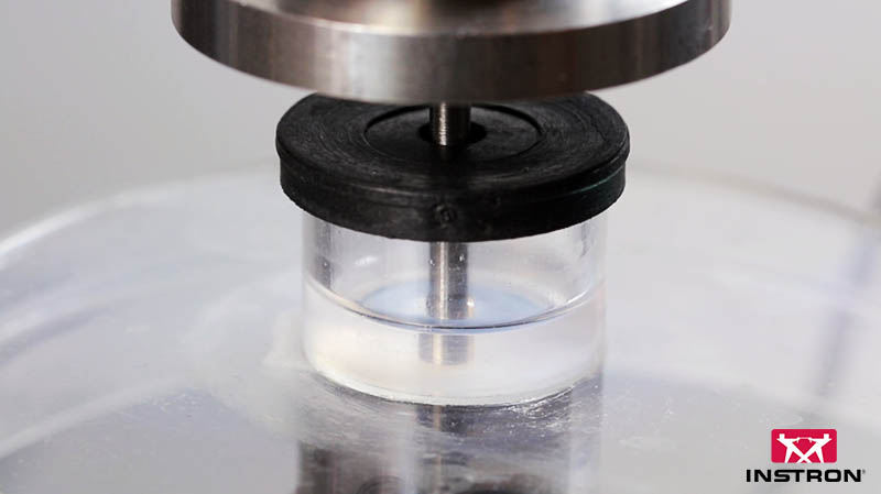
The Instron ElectroPuls E3000 was fitted with a 50N load cell. 150μm-thick collagen gels were placed into a fluid-filled petri dish container. Initially, a simple waveform was created with a 0.05 μm/s indentation rate.
Since it was difficult to accurately place the indenter a few μm above the surface of the gel, tests ran for a very long time. A more complex waveform was created that would indent the gel up to 0.5N, then raise the indenter 150μm to just above the gel surface. This reduced indentation times by a factor of 4 while still maintaining 0.05 μm/s indentation rates.
This ultra-slow indentation allowed for precise mapping of the force-indentation profile of the thin gel, from which stiffness could be estimated.
Results were processed using a low-pass filter to further reduce noise, along with a downsample filter to reduce file size. An algorithm using linear modeling analysis found the start point of the test, as well as the point where plastic deformation beneath the surface became significant.
From the load-depth profile, the modulus of the collagen gels was calculated using the following formula:
Where F is the load applied, E is the elastic modulus, R is the radius of the indenter, δ is the indentation depth and ν is Poisson’s ratio of the material.
The Instron Advantage
Testing thin film tissue samples with the ElectroPuls E3000 — on the same system used for dynamic 3 kN fatigue tests — demonstrates the flexibility of the instrument, with the limits of what the machine can do pushed further by the ingenuity of the user. Instron® offers a full range of load cells suited to a wide range of different applications.
Since modulus values depend on the speed of loading, accurate modulus values for biomechanical modeling need to come from tests run at physiological speeds. The ElectroPuls' high-fidelity control loops for positional accuracy allow it to perform creep-style tests on the order of micrometers per minute, approaching physiological speeds.
Because post-processing gives the user access to raw data, end users can process that data however they see fit, without integral system biases.
The calculations module's ability to enhance and expand the machine's capabilities was demonstrated by the noise reduction technique used. A simple noise reduction algorithm, which takes the mean average of all incoming data, shows how the system can be tuned to whatever test is needed using the calculation module.
This approach allowed information from 10 kHz of data to be used while only recording data at 10 Hz, keeping file sizes manageable while significantly reducing noise levels — all through the addition of a single calculation.
Results
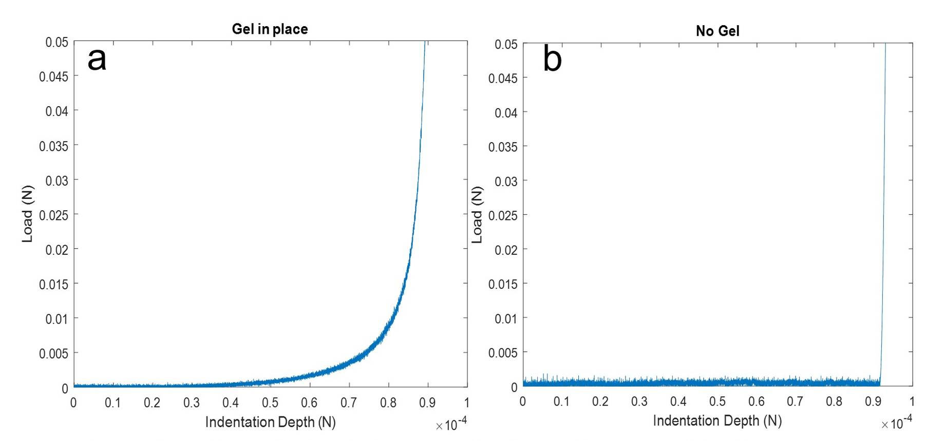
The two graphs below show that a load cell is capable of providing mechanical information from soft tissue as thin as a 150μm collagen gel.
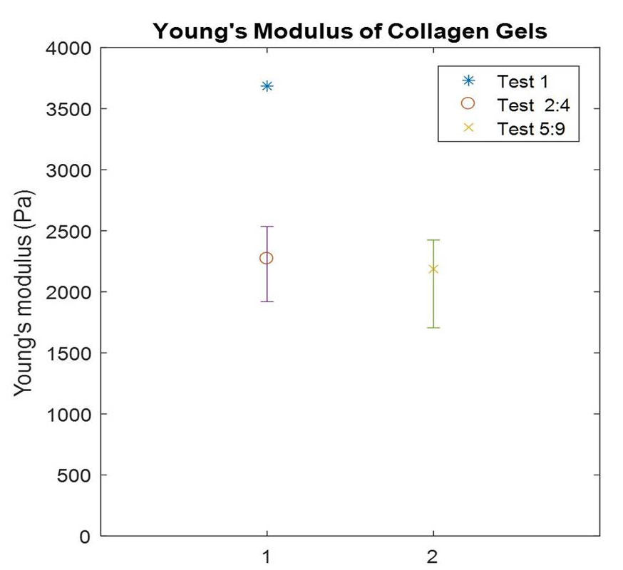
Final results show that the modulus of collagen in this form falls in the range of 1800 Pa to 2500 Pa (Figure 2). These results can be incorporated into models of tumor growth, and they also demonstrate that meaningful results can be obtained from these gels — meaning future research will be able to characterize collagen-embedded cancer cells to build a better model of cancer growth.
These results were validated using an atomic force microscope (AFM), typically used for modulus measurements on much smaller samples. The AFM results agreed with those from the ElectroPuls™, suggesting that for this type of testing, ElectroPuls™ has the capability and sensitivity to test thin biological tissues.
Acknowledgments
Many thanks to:

Talk to an Instron Expert.
Not sure if the ElectroPuls E3000 fits your low-force or biomedical testing application? Tell us about your requirements and we will help you find the right solution.
Talk to an Applications Engineer
Tell us about your testing needs, and we'll help you configure the right frame, load cell, grips, and software for your lab. Please complete the form below, and we'll respond within 1-2 business days. For a faster response, call +1-800-473-7838 or +1 781 828 2500 if you are outside of the US.